OVERVIEW
Spina bifida occurs during the third and fourth weeks of pregnancy when a portion of the fetal spinal cord fails to properly close. As a result, the child is born with a part of the spinal cord exposed on the back.
Causes
Spina bifida is the most common, permanently disabling birth defect. In 2005, the CDC approximated that about 18 cases of myelomeningocele per 100,000 live births occur in the United States. The Spina Bifida Association conservatively estimates that there are 70,000 people living in the United States with the condition. The prevalence appears to have decreased in recent years due in part to preventative measures followed by expectant mothers prior to and during pregnancy as well as prenatal testing. The risk of having a second affected child increases to 2-3% and 10% for a third child.
Although scientists believe that genetic and environmental factors may act together to cause spina bifida, 95% of babies with spina bifida are born to parents with no family history. Women with certain chronic health problems, including diabetes and seizure disorders (treated with certain anticonvulsant medications), have an increased risk (approximately 1/100) of having a baby with spina bifida.
Symptoms
The symptoms of spina bifida depend on exactly where and what extent the spinal cord and overlying structures have not developed correctly. There are three common subtypes:
Occulta is often called hidden spina bifida, as the spinal cord and the nerves are usually normal and there is no opening on the back. In this form of spina bifida, there is only a small defect or gap in the small bones (vertebrae) that make up the spine, which occurs in about 12% of the population. In many cases, spina bifida occulta is so mild that there is no disturbance of spinal function at all. Most people are not aware that they have spina bifida occulta, unless it is discovered on an x-ray performed for an unrelated reason. However, one in 1,000 individuals will have an occult structural finding that leads to neurological deficits or disabilities as bowel or bladder dysfunction, back pain, leg weakness or scoliosis.
Meningocele occurs when the bones do not close around the spinal cord and the meninges are pushed out through the opening, causing a fluid-filled sac to form. The meninges are three layers of membranes covering the spinal cord, consisting of dura mater, arachnoid mater and pia mater. In most cases, the spinal cord and the nerves themselves are normal or not severely affected. The sac is often covered by skin and may require surgery. This is the rarest type of spina bifida.
Myelomeningocele accounts for about 75% of all spina bifida cases. This is the most severe form of the condition in which a portion of the spinal cord itself protrudes through the back. In some cases, sacs are covered with skin, but in other cases, tissue and nerves may be exposed. The extent of neurological disabilities is directly related to the location and severity of the spinal cord defect. If the bottom of the spinal cord is involved, there may be only bowel and bladder dysfunction, while the more severe cases can result in total paralysis of the legs with accompanying bowel and bladder dysfunction.
When & How to Seek Medical Care
The defect is most often detected on maternal screening tests and/or ultrasound of the fetus. Babies whose mother’s lack access to care or those with extremely small lesions may not be diagnosed until after delivery.
When this type of defect is detected, parents should seek out a multi-disciplinary team of specialists, including a pediatric neurosurgeon, to understand the condition and prognosis as well as to make an informed decision on the course of the pregnancy, possible further fetal testing, type of delivery and consideration of fetal surgery.
Testing & Diagnosis
- Maternal serum alpha-fetoprotein test (Physican may order follow-up tests for confirmation including amniocentesis)
- 20-week ultrasound
- Infant ultrasound
- Fetal MRI
Treatment
Pre-natal
Women of childbearing age can reduce their risk of having a child with spina bifida by taking 400 micrograms (mcg) of folic acid every day pre-conception. Because it is water soluble, folic acid does not stay in the body for very long and needs to be taken every day to be effective against neural defects. Since half of all pregnancies in the United States are unplanned, folic acid must be taken whether a woman is planning a pregnancy or not. Research has shown that if all women of childbearing age took a multivitamin with the B-vitamin folic acid, the risk of neural tube defects could be reduced by up to 70%.
Fetal Surgery
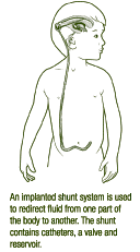
A randomized study (MOMS Trial) compared repair of the defect while the fetus was still in the uterus versus repair after birth and had encouraging, but mixed, results. Patients who underwent fetal repair were both less likely to need a ventriculo-peritoneal shunt (44 vs. 84%) and more likely to be able to walk (44 vs. 24%). The fetal surgery group also had many more maternal and infant complications. Given the higher risk of complications as well as the lack of long-term follow-up, the current recommendation of the American College of Obstetricians and Gynecologists (ACOG) is that fetal surgery only be considered at specialty centers with teams experienced in fetal surgery.
Follow-up
Each patient should have baseline imaging as well as detailed neurologic and muscular exams. The most common causes of deterioration are often treatable and therefore robust reference point and longitudinal assessments are necessary to identify them in a timely manner.
Outlook
If a child is paraplegic (has no movement in the legs from the hips down), he or she will need a wheelchair. If a child is born with movement of the thigh muscles and feeling down to below the knees, the chances are good he or she will be able to walk with some sort of brace support. When there are no brain abnormalities, the child may have average or above average intelligence, even if there is advanced hydrocephalus at birth.
Fortunately, with proper medical care, many children with spina bifida can lead active and productive lives. Twenty-year follow-up studies of children with spina bifida show that they enter college in the same proportion as the general population and many are actively employed. With recent advancements in medical care for these children, their outlook continues to improve.

RESOURCES
Note from AANS
The AANS does not endorse any treatments, procedures, products or physicians referenced in these patient fact sheets. This information provided is an educational service and is not intended to serve as medical advice. Anyone seeking specific neurosurgical advice or assistance should consult his or her neurosurgeon, or locate one in your area through the AANS’ Find a Board-certified Neurosurgeon online tool.
